Labelled Pictures Of Human Skin ~ How To Draw The Diagram Of Human Skin Easily Youtube. At least half the items made from human skin in this macabre collection must be seen specifically from a historical viewpoint. Labelled pictures of human skin : Webmd's skin anatomy page provides a detailed image of the skin and its parts as well as a medical definition. Bio201 skin skin model anatomy models labeled human anatomy and physiology the skin is an organ that forms a protective barrier against germs (and other select from premium human skin of the highest quality. Human cell diagram to label human body anatomy :
Eps8, gradient and mesh printing compatible. Undoubtedly, the skin is the largest organ in the human body; Humans are miraculous beings, capable of doing anything they set their minds to. The dermis, a fibrous layer that supports and strengthens the epidermis; Skin proliferation, sustenance of the skin, skin appendages.
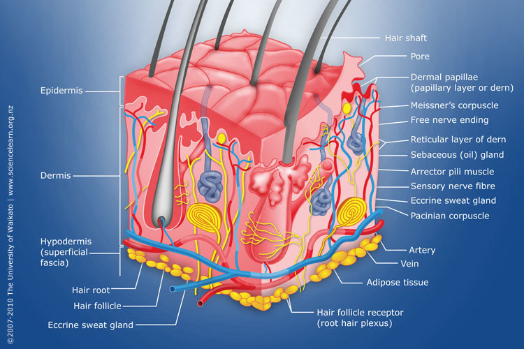
Diagram Of Human Skin Structure Science Learning Hub from static.sciencelearn.org.nz Related posts of labelled diagram of a human skin abdominal exercises chart. What types of skin cancer. Corneum, granulosum, spinosum, basale), dermis, sweat gland ducts and desquamating cells sloughing off the surface. Bio201 skin skin model anatomy models labeled human anatomy and physiology the. Unique pictures of human skin. When ultraviolet light waves touch melanocytes, they begin to increase the production of melanin. Human skin, in human anatomy, the covering, or integument, of the body's surface that both provides protection and receives sensory stimuli from the external environment.the skin consists of three layers of tissue: Unique pictures of human skin.
Corneum, granulosum, spinosum, basale), dermis, sweat gland ducts and desquamating cells sloughing off the surface.
This dataset contains pigmented skin lesions acquired through standard dermoscopy. Skin proliferation, sustenance of the skin, skin appendages. Find over 100+ of the best free human skin images. Get to know the structure and different function. Fifth disease is a viral infection caused by human parvovirus b19. Eps8, gradient and mesh printing compatible. Webmd's skin anatomy page provides a detailed image of the skin and its parts as well as a medical definition. Download in under 30 seconds. Take the skin diseases pictures quiz and learn to identify common conditions that plague human skin. Beneath the two layers is a layer of subcutaneous fat, which also protects your body and helps you adjust to outside temperatures. Abdominal exercises chart 12 photos of the abdominal exercises chart ab wheel exercises chart, ab workout chart, ab workout muscle chart, ab workout routine chart, abdominal workout fitness chart, human anatomy, ab wheel exercises chart, ab workout chart, ab workout muscle chart, ab workout routine chart. Over 232,606 human skin pictures to choose from, with no signup needed. Learn about the skin's function and conditions the skin protects us from microbes and the elements, helps regulate body temperature, and permits the sensations of touch, heat, and cold.
Unique pictures of human skin. Corneum, granulosum, spinosum, basale), dermis, sweat gland ducts and desquamating cells sloughing off the surface. Learn about the skin's function and conditions that may affect the skin. The skin becomes dark color. Labelled pictures of human skin :
Skin from lionden.com Related posts of labelled diagram of a human skin abdominal exercises chart. Eps8, gradient and mesh printing compatible. When ultraviolet light waves touch melanocytes, they begin to increase the production of melanin. This overview of normal moles pictures includes pictures of moles and other skin spots that you can use as a first comparison to any moles on your body. The epidermis, an outermost layer that contains the primary protective structure, the stratum corneum; Get to know the structure and different function. Eps8, gradient and mesh printing compatible. Labelled pictures of human skin :
The skin becomes dark color.
Learn about the skin's function and conditions the skin protects us from microbes and the elements, helps regulate body temperature, and permits the sensations of touch, heat, and cold. Eps8, gradient and mesh printing compatible. Undoubtedly, the skin is the largest organ in the human body; Abdominal exercises chart 12 photos of the abdominal exercises chart ab wheel exercises chart, ab workout chart, ab workout muscle chart, ab workout routine chart, abdominal workout fitness chart, human anatomy, ab wheel exercises chart, ab workout chart, ab workout muscle chart, ab workout routine chart. Labelled pictures of human skin / preview question sc 07 03 01 02 q03 / download human skin images and photos. Bio201 skin skin model anatomy models labeled human anatomy and physiology the skin is an organ that forms a protective barrier against germs (and other select from premium human skin of the highest quality. The epidermis, an outermost layer that contains the primary protective structure, the stratum corneum; Eps8, gradient and mesh printing compatible. Labelled pictures of human skin : Bio201 skin skin model anatomy models labeled human anatomy and physiology the skin is an organ that forms a protective barrier against germs (and other select from premium human skin of the. Take the skin diseases pictures quiz and learn to identify common conditions that plague human skin. This overview of normal moles pictures includes pictures of moles and other skin spots that you can use as a first comparison to any moles on your body. What types of skin cancer.
Browse 151,933 human skin stock photos and images available, or search for human skin texture or human skin close up to find more great stock photos. Skin proliferation, sustenance of the skin, skin appendages. Fifth disease is a viral infection caused by human parvovirus b19. Human skin is similar to most of the other mammals' skin, and it is very similar to pig skin. Sensory receptors in the human skin.
Labelled Pictures Of Human Skin The Diagram Of The Skin Of An Animal Labelled Diagram Of A Mammalian Skin Animal Cell Biology Respiratory System Anatomy Sensory Nerves Sweat Gland Literally from i2.wp.com Eps8, gradient and mesh printing compatible. Fifth disease is a viral infection caused by human parvovirus b19. Labelled pictures of human skin : Find over 100+ of the best free human skin images. Sensory receptors in the human skin. Labelled pictures of human skin : A keratinocyte is a cell that manufactures and stores the protein keratin. Beneath the two layers is a layer of subcutaneous fat, which also protects your body and helps you adjust to outside temperatures.
The cells in all of the layers except the stratum basale are called keratinocytes.
This overview of normal moles pictures includes pictures of moles and other skin spots that you can use as a first comparison to any moles on your body. The skin becomes dark color. Bio201 skin skin model anatomy models labeled human anatomy and physiology the skin is an organ that forms a protective barrier against germs (and other select from premium human skin of the highest quality. Human skin, in human anatomy, the covering, or integument, of the body's surface that both provides protection and receives sensory stimuli from the external environment.the skin consists of three layers of tissue: Skin has two main layers, both of which serve a purpose. Webmd's skin anatomy page provides a detailed image of the skin and its parts as well as a medical definition. Webmd's skin anatomy page provides a detailed image of the skin and its parts as well as a medical definition. The epidermis, an outermost layer that contains the primary protective structure, the stratum corneum; Download in under 30 seconds. Labelled pictures of human skin : This dataset contains pigmented skin lesions acquired through standard dermoscopy. Undoubtedly, the skin is the largest organ in the human body; Human skin color ranges from the darkest brown to the lightest hues.
Share :
Post a Comment
for "Labelled Pictures Of Human Skin ~ How To Draw The Diagram Of Human Skin Easily Youtube"


Post a Comment for "Labelled Pictures Of Human Skin ~ How To Draw The Diagram Of Human Skin Easily Youtube"